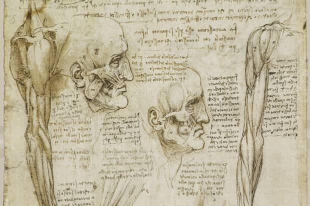
The Queen’s Gallery at Buckingham Palace will host the largest ever display of Leonardo da Vinci’s anatomical drawings in the exhibition “Leonardo da Vinci: Anatomist” through the 7th of October 2012.
Showcasing 87 pages from da Vinci’s notebooks, the exhibit included 24 drawings that have never before been publically displayed. Several of da Vinci’s early anatomical drawings, including “The Anatomy of a Bear’s Foot” and “A Skull Sectioned” from the late 1480s, will be displayed, as well as detailed sketches from his human dissection period, notably the first accurate representation of the spine, studies on the valves of the heart, and his famous, though not entirely accurate, chalk drawing of a foetus in the womb.
After da Vinci’s death in 1519, the significance of his anatomical work went unnoticed, as it was so far ahead of medical knowledge during that period. In 1543, French anatomist Andreas Vesalius published his treatise De humani corporis fabrica (The Fabric of the Human Body), which, though it contained fewer discoveries than da Vinci’s drawings, became the most important anatomical work ever published. Some of the anatomic features described by da Vinci were not confirmed until MRI imaging studies 30 years ago.
Exhibition curator Martin Clayton told ArtDaily that “had [da Vinci] published this work, he would now be known as one of the greatest scientists in history.” Though the drawings have been in the Royal Collection since at least 1690, their significance was not noted until the 20th century. Anatomists now believe that many of da Vinci’s drawings are as accurate anything produced by scientific artists today and are superseded in quality only by digital technology.
An iPad app accompanying the exhibit includes all 268 of da Vinci’s extant anatomical drawings from the Royal Collection, as well as a feature that reverses and translates da Vinci’s notes, rendering them accessible to the public. The drawings included in the exhibit are also available on the Royal Collection website.


